
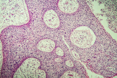
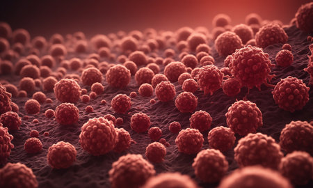
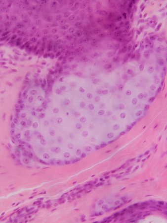
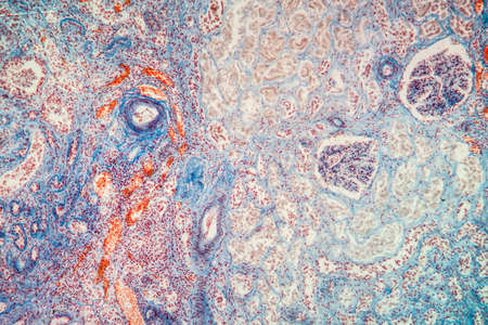
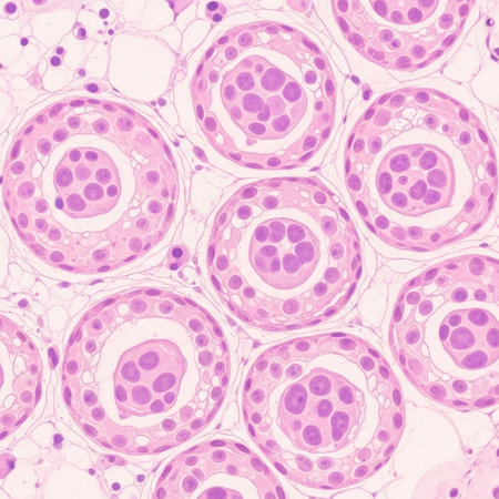
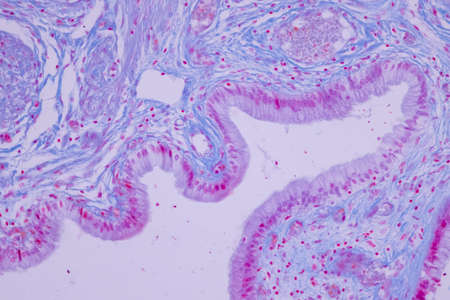
Bowen's Disease Tumor under the microscope 100x
Коллекция по умолчанию
Коллекция по умолчанию
Создать новую
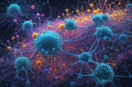
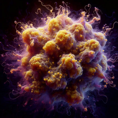
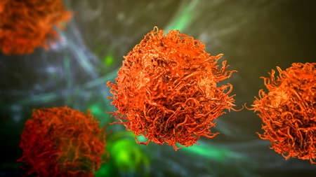
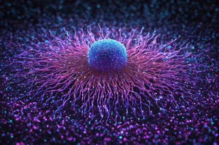
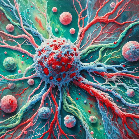
Cosmic Neural Network. Generative AI.
Коллекция по умолчанию
Коллекция по умолчанию
Создать новую
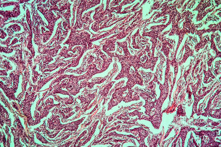
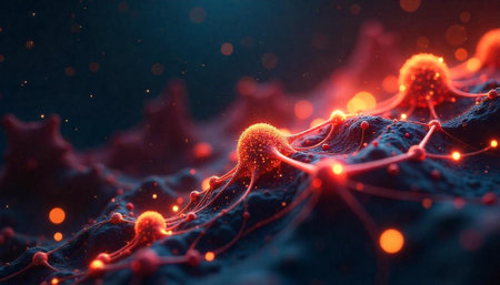
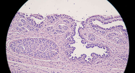
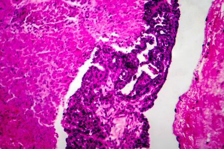
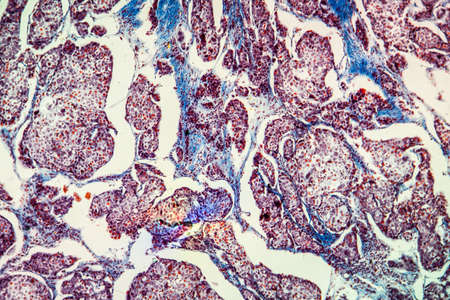
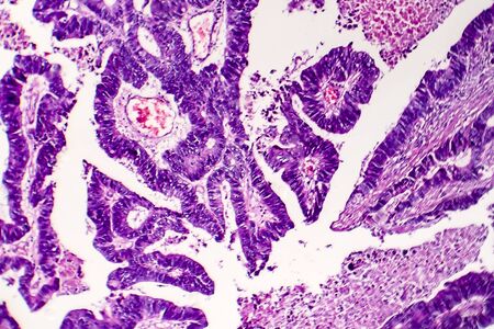
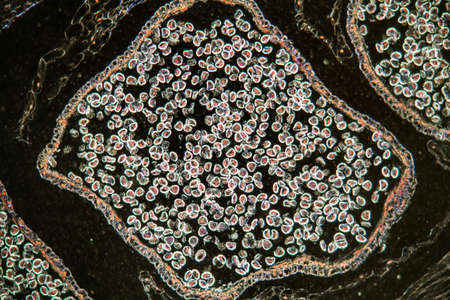
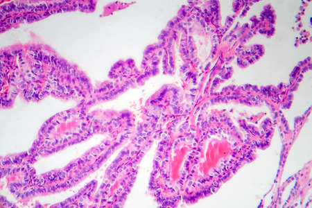
Breast cancer of the woman diseased tissue 100x
Коллекция по умолчанию
Коллекция по умолчанию
Создать новую
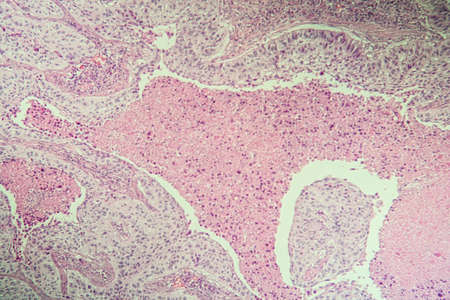
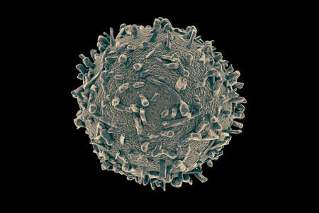
Squamous cell carcinoma diseased tissue under the microscope 100x
Коллекция по умолчанию
Коллекция по умолчанию
Создать новую
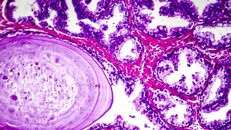
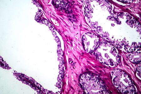
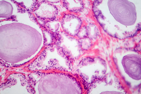
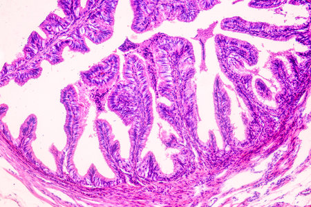
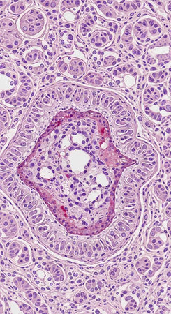
Histopathology of prostate gland hyperplasia, light micrograph, photo under microscope
Коллекция по умолчанию
Коллекция по умолчанию
Создать новую

Jellyfish swimming in the water. 3D illustration. Jellyfish macro
Коллекция по умолчанию
Коллекция по умолчанию
Создать новую
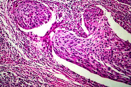
Squamous cell carcinoma of the uterus, light micrograph, photo under microscope
Коллекция по умолчанию
Коллекция по умолчанию
Создать новую
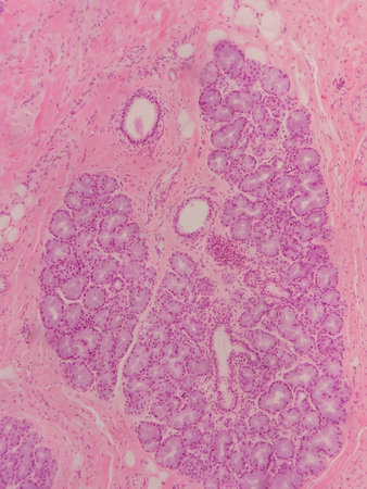
Salivary gland swollen diseased tissue under the microscope 100x
Коллекция по умолчанию
Коллекция по умолчанию
Создать новую
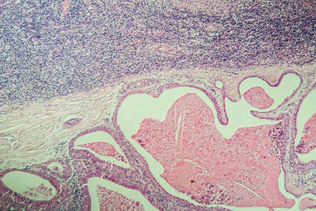
Light micrograph of teratoma, a tumor made up of several different types of tissue, such as hair, teeth, muscle, or bone. Teratoma is typically found in the ovary, testicle, or coccyx
Коллекция по умолчанию
Коллекция по умолчанию
Создать новую
fish caviar as a background. macro
Коллекция по умолчанию
Коллекция по умолчанию
Создать новую
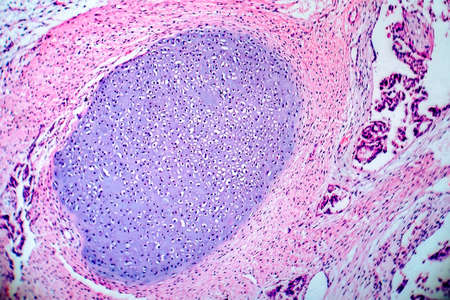
Condyloma acuminatum, also known as genital warts. Light micrograph, photo under microscope
Коллекция по умолчанию
Коллекция по умолчанию
Создать новую

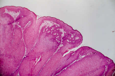
Bowen's Disease Tumor under the microscope 100x
Коллекция по умолчанию
Коллекция по умолчанию
Создать новую
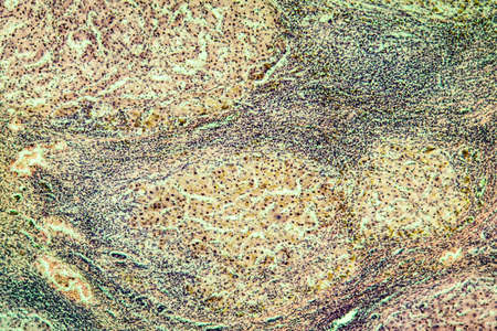
Liver cirrhosis tissue affected 100x after alcohol abuse
Коллекция по умолчанию
Коллекция по умолчанию
Создать новую
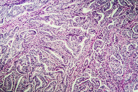
Gastric carcinoma in tissue section 100x
Коллекция по умолчанию
Коллекция по умолчанию
Создать новую
Bacteria, Bacterial colony, Microbes.
Коллекция по умолчанию
Коллекция по умолчанию
Создать новую

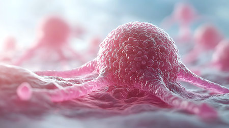
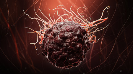
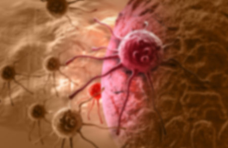
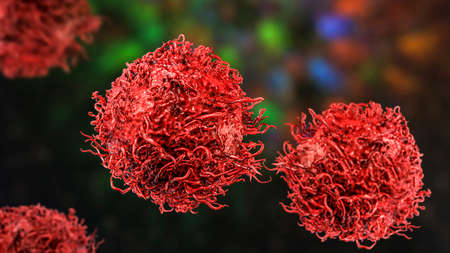
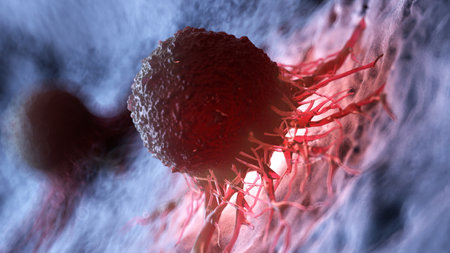
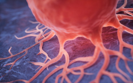
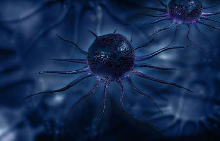
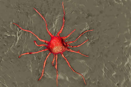
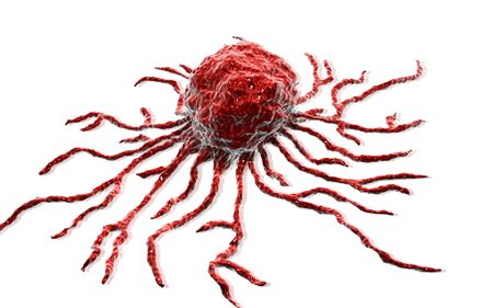
Cancer cell, malignant tumor cell
Коллекция по умолчанию
Коллекция по умолчанию
Создать новую

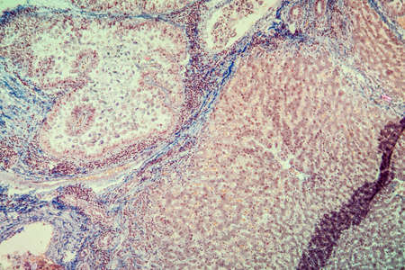
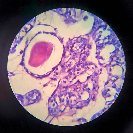
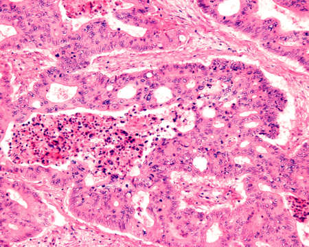
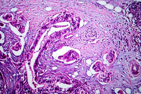
Ovarian cancer, light micrograph, photo under microscope. Photograph shows a fragment of a cancerous tumor in the female ovary. Selective focus
Коллекция по умолчанию
Коллекция по умолчанию
Создать новую
A biohazard abstraction, Ai generated image
Коллекция по умолчанию
Коллекция по умолчанию
Создать новую
Interconnected network of cells or organisms with glowing connections
Коллекция по умолчанию
Коллекция по умолчанию
Создать новую
Prostate cancer, light micrograph, photo under microscope
Коллекция по умолчанию
Коллекция по умолчанию
Создать новую
cancer cell or tumor illustration in high detail for medical concept 3d rendering
Коллекция по умолчанию
Коллекция по умолчанию
Создать новую
3d rendered illustration of a cancer cell
Коллекция по умолчанию
Коллекция по умолчанию
Создать новую
Histopathology of human liver under microscope view for medical education.
Коллекция по умолчанию
Коллекция по умолчанию
Создать новую
Coccidiosis of liver tissue under the microscope 100x
Коллекция по умолчанию
Коллекция по умолчанию
Создать новую
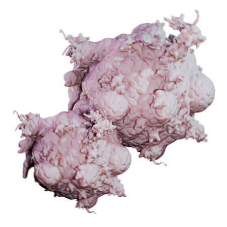
tumor mass isolated on white background 3d illustration
Коллекция по умолчанию
Коллекция по умолчанию
Создать новую
Thyroid follicular carcinoma, light micrograph, photo under microscope
Коллекция по умолчанию
Коллекция по умолчанию
Создать новую
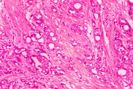
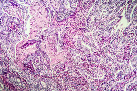
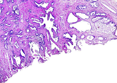
Histopathology of adenocarcinoma of the prostate
Коллекция по умолчанию
Коллекция по умолчанию
Создать новую
Colon inflammation in Crohn's disease 100x
Коллекция по умолчанию
Коллекция по умолчанию
Создать новую
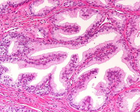
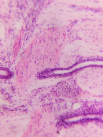
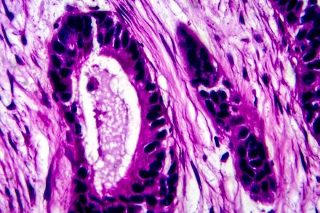
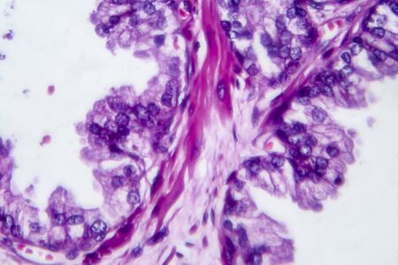
High magnification of a human prostatic gland. A simple columnar epithelium surrounds a very irregular lumen. Hematoxylin & eosin stain.
Коллекция по умолчанию
Коллекция по умолчанию
Создать новую
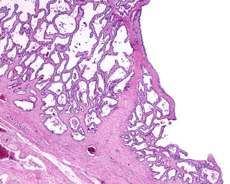
Photomicrograph showing histological features of benign prostatic hyperplasia. Enlarged prostate gland with nodular proliferation of glandular and stromal components.
Коллекция по умолчанию
Коллекция по умолчанию
Создать новую
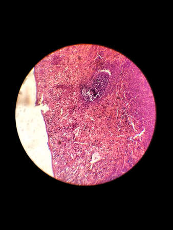
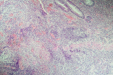
Biological histological fixed colored preparation of the spleen - a secondary organ of the immune system
Коллекция по умолчанию
Коллекция по умолчанию
Создать новую
Anatomy and Histological Ovary, Testis and Sperm human cells under microscope.
Коллекция по умолчанию
Коллекция по умолчанию
Создать новую
Human liver tissue under microscope view. Histological sample of human liver.
Коллекция по умолчанию
Коллекция по умолчанию
Создать новую
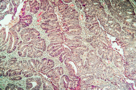
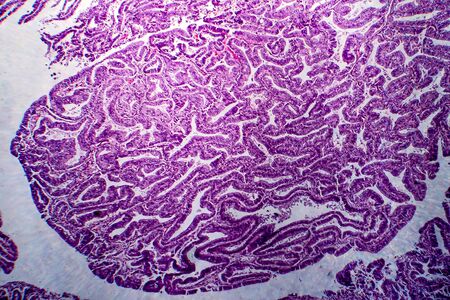
Papillary serous ovarian adenocarcinoma, cancer of ovary, light micrograph, photo under microscope
Коллекция по умолчанию
Коллекция по умолчанию
Создать новую
Photomicrograph showing histological features of benign prostatic hyperplasia. Enlarged prostate gland with nodular proliferation of glandular and stromal components.
Коллекция по умолчанию
Коллекция по умолчанию
Создать новую
Cancer or tumor cell close-up 3D rendering illustration. Carcinoma, lymphoma, oncology, medicine, science, microbiology, cancerous pathology, health concepts.
Коллекция по умолчанию
Коллекция по умолчанию
Создать новую

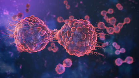
Cancer cells mitosis or proliferation 3D rendering illustration. Division of two malignant cells causing carcinoma close-up. Medicine, oncology, science, disease, biology and microbiology concepts.
Коллекция по умолчанию
Коллекция по умолчанию
Создать новую
bacteria cells and virus under the microscope, concept of microbiology, scientific and medical research (3d render)
Коллекция по умолчанию
Коллекция по умолчанию
Создать новую

fantasy diagram of a interior of a cell or microorganism is presented, including the cell wall, cytoplasm, and distinct organelles like the nucleus, mitochondria, vacuole, and endoplasmic reticulum.
Коллекция по умолчанию
Коллекция по умолчанию
Создать новую
cancer cell colon made in 3d software
Коллекция по умолчанию
Коллекция по умолчанию
Создать новую
Basal cell cancer Diseased tissue 100x
Коллекция по умолчанию
Коллекция по умолчанию
Создать новую
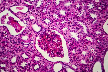
Chronic pyelonephritis, light micrograph, photo under microscope
Коллекция по умолчанию
Коллекция по умолчанию
Создать новую
A microscopic view of tissue with pink and purple staining, showing cellular structures and patterns
Коллекция по умолчанию
Коллекция по умолчанию
Создать новую
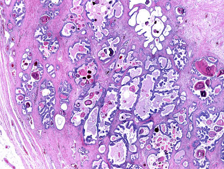
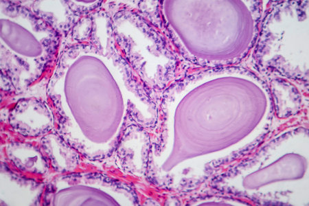
Low magnification of a human prostate gland in a 70-year-old man. The prostate gland appears with dilated alveoli, which contains many corpora amylacea (prostatic concretions) in their lumen. Light microscope micrograph. Hematoxylin & eosin stain.
Коллекция по умолчанию
Коллекция по умолчанию
Создать новую
Condyloma acuminatum, also known as genital warts. Light micrograph, photo under microscope
Коллекция по умолчанию
Коллекция по умолчанию
Создать новую
Stomach cancer cells, 3D illustration showing morphology of cancerous cells
Коллекция по умолчанию
Коллекция по умолчанию
Создать новую
Esophageal squamous cell carcinoma, light micrograph, photo under microscope
Коллекция по умолчанию
Коллекция по умолчанию
Создать новую
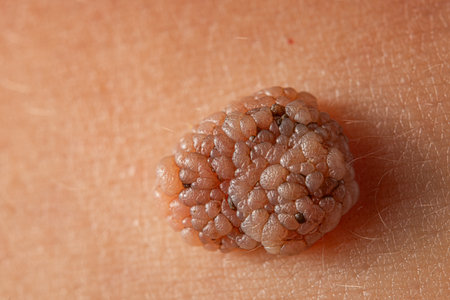
Close-up of a large mole on the body of a caucasian man. macro shot.
Коллекция по умолчанию
Коллекция по умолчанию
Создать новую

Bladder transitional cell carcinoma, light micrograph, photo under microscope
Коллекция по умолчанию
Коллекция по умолчанию
Создать новую
Uterine cancer, light micrograph, photo under microscope
Коллекция по умолчанию
Коллекция по умолчанию
Создать новую
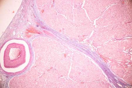
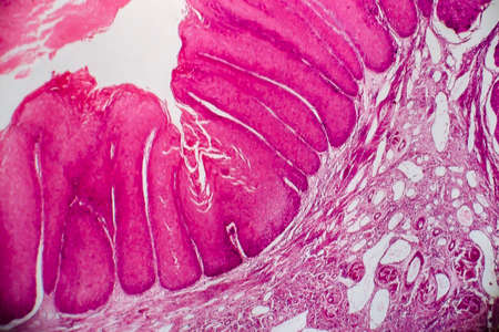
Human seminal vesicle. The surface of the mucosa is very folded. The spaces that look like glands are really infoldings of the mucosa that communicate with the lumen. The epithelium is pseudostratified columnar with basal cells.
Коллекция по умолчанию
Коллекция по умолчанию
Создать новую
3d rendered illustration of a human cancer cell
Коллекция по умолчанию
Коллекция по умолчанию
Создать новую
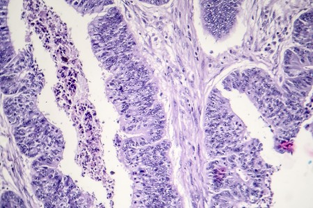
Large-bowel adenocarcinoma. Cancer cells arranged in cords or strands with empty central spaces remembering the normal crypts of the colon mucosa.
Коллекция по умолчанию
Коллекция по умолчанию
Создать новую
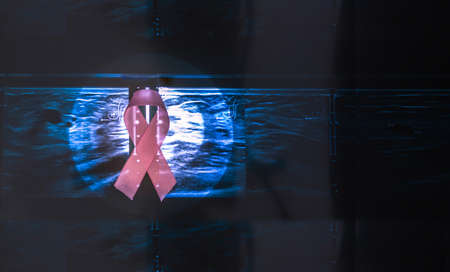
Pink breast cancer awareness ribbon on a mammography background Breast cancer prevention concept
Коллекция по умолчанию
Коллекция по умолчанию
Создать новую
Colon carcinoma arising from adenoma, 100x
Коллекция по умолчанию
Коллекция по умолчанию
Создать новую
Stomach cancer cells, 3D illustration showing morphology of cancerous cells
Коллекция по умолчанию
Коллекция по умолчанию
Создать новую
Microscopic view of tissue with purple and pink staining, showing cellular structures and nuclei
Коллекция по умолчанию
Коллекция по умолчанию
Создать новую
Differentiated intestinal adenocarcinoma, light micrograph, photo under microscope
Коллекция по умолчанию
Коллекция по умолчанию
Создать новую

3d rendered illustration of human cancer cells
Коллекция по умолчанию
Коллекция по умолчанию
Создать новую

Lung adenocarcinoma, light micrograph, photo under microscope
Коллекция по умолчанию
Коллекция по умолчанию
Создать новую
White blood cells (WBCs), also called leukocytes or leucocytes, are the cells of the immune system that are involved in protecting the body against both infectious disease and foreign invaders. (WIkipedia)
Коллекция по умолчанию
Коллекция по умолчанию
Создать новую

Histopathology of interstitial nephritis, light micrograph, photo under microscope
Коллекция по умолчанию
Коллекция по умолчанию
Создать новую
Prostate cancer, light micrograph, photo under microscope
Коллекция по умолчанию
Коллекция по умолчанию
Создать новую
Gastric carcinoma in tissue section 100x
Коллекция по умолчанию
Коллекция по умолчанию
Создать новую
A biohazard abstraction, Ai generated image
Коллекция по умолчанию
Коллекция по умолчанию
Создать новую

Kidney cancer, light micrograph, photo under microscope. High magnification
Коллекция по умолчанию
Коллекция по умолчанию
Создать новую

Uterine cancer, light micrograph, photo under microscope
Коллекция по умолчанию
Коллекция по умолчанию
Создать новую
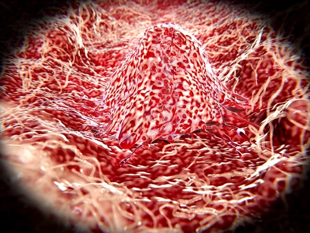
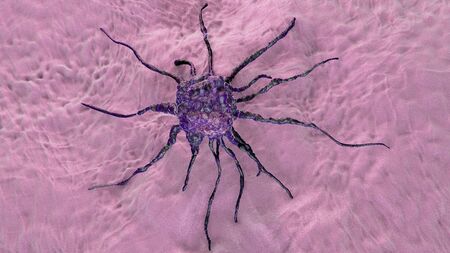
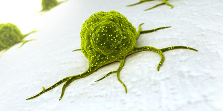
Migrating cancer cell
Коллекция по умолчанию
Коллекция по умолчанию
Создать новую
Digital illustration of stem cell in color background
Коллекция по умолчанию
Коллекция по умолчанию
Создать новую
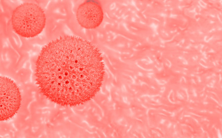
Illustration of a virus cell in a vein.
Коллекция по умолчанию
Коллекция по умолчанию
Создать новую
Biological cancer cell and disease, 3d rendering. 3D illustration.
Коллекция по умолчанию
Коллекция по умолчанию
Создать новую
Breast cancer, light micrograph, photo under microscope
Коллекция по умолчанию
Коллекция по умолчанию
Создать новую

Colon polyp, one of the largest polyps
Коллекция по умолчанию
Коллекция по умолчанию
Создать новую

3d illustration CAR T-cell attack cancer cell
Коллекция по умолчанию
Коллекция по умолчанию
Создать новую

Abstract science background- pyloric division of the stomach of the dog. Cell biology
Коллекция по умолчанию
Коллекция по умолчанию
Создать новую

ancient plankton organism
Коллекция по умолчанию
Коллекция по умолчанию
Создать новую
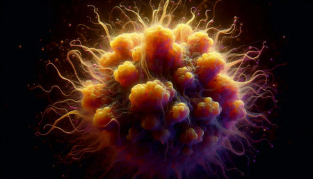
A holographic the microbe, made up of many particles. Generative AI.
Коллекция по умолчанию
Коллекция по умолчанию
Создать новую
Digital illustration of stem cell in color background
Коллекция по умолчанию
Коллекция по умолчанию
Создать новую
Education anatomy and Histological sample of Human under the microscope.
Коллекция по умолчанию
Коллекция по умолчанию
Создать новую

close-up coronavirus type covid-19, h1n1, bird flu or swine flu background. 3d rendering of representation of virus as microbiological microscopic background. Red white gradient color
Коллекция по умолчанию
Коллекция по умолчанию
Создать новую
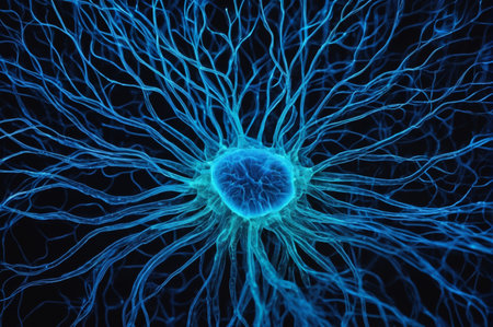
Neural Core Network. Generative AI.
Коллекция по умолчанию
Коллекция по умолчанию
Создать новую

Bladder cat- cell nature background. Abstract- photo macro sections with high magnification with light microscope
Коллекция по умолчанию
Коллекция по умолчанию
Создать новую
Atrophy kidney tissue under the microscope 100x
Коллекция по умолчанию
Коллекция по умолчанию
Создать новую
Cancer cell, tumour cell, close-up view, 3D illustration
Коллекция по умолчанию
Коллекция по умолчанию
Создать новую
Cancer cell, tumour cell, close-up view, 3D illustration
Коллекция по умолчанию
Коллекция по умолчанию
Создать новую
Club moss flower with seeds under the microscope 100x
Коллекция по умолчанию
Коллекция по умолчанию
Создать новую
Pancreas cancer, light micrograph, photo under microscope
Коллекция по умолчанию
Коллекция по умолчанию
Создать новую
Digital illustration of stem cell in isolated background
Коллекция по умолчанию
Коллекция по умолчанию
Создать новую
3d rendered illustration of a cancer cell
Коллекция по умолчанию
Коллекция по умолчанию
Создать новую
Papillary thyroid carcinoma, light micrograph, photo under microscope. The most common type of thyroid cancer
Коллекция по умолчанию
Коллекция по умолчанию
Создать новую
A holographic Digital molecule of a substance, made up of many particles. Generative AI.
Коллекция по умолчанию
Коллекция по умолчанию
Создать новую
Histopathology of fibroids, light micrograph, photo under microscope
Коллекция по умолчанию
Коллекция по умолчанию
Создать новую

Endemic goiter, light micrograph, abnormal enlargement of the thyroid gland due to dietary iodine deficiency. Photomicrograph shows follicles of varying size, abundant colloid, lymphocytic infiltrate
Коллекция по умолчанию
Коллекция по умолчанию
Создать новую
Endometrial adenocarcinoma, light micrograph, photo under microscope
Коллекция по умолчанию
Коллекция по умолчанию
Создать новую
Prostate cancer of a human, highly detailed segment of panorama. Photomicrograph as seen under the microscope, 10x zoom.
Коллекция по умолчанию
Коллекция по умолчанию
Создать новую

Hard breast cancer, light micrograph, photo under microscope
Коллекция по умолчанию
Коллекция по умолчанию
Создать новую

Doctor measuring size of patients pigmented nevus with ruler in clinic closeup. Diagnosis of skin tumors concept
Коллекция по умолчанию
Коллекция по умолчанию
Создать новую

Fantasy of pink milky way galaxy with stars and darkness space background in romance valentine concept.
Коллекция по умолчанию
Коллекция по умолчанию
Создать новую
Columnar epithelium of human gall bladder under the microscope in Lab.
Коллекция по умолчанию
Коллекция по умолчанию
Создать новую
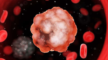
Enlarged image of cancer cells, tumors, blood cancers
Коллекция по умолчанию
Коллекция по умолчанию
Создать новую
Legion-Media
Создайте свои проекты на основе качественных стоковых фотографий и видео.


Copyright © Legion-Media.